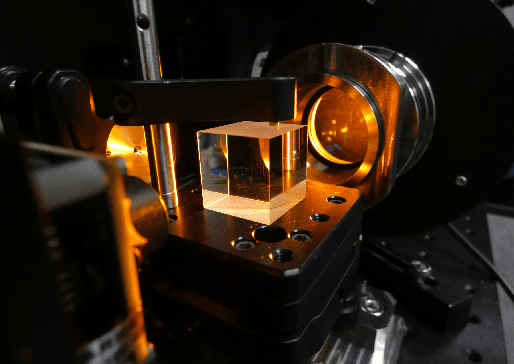
Optical microscopy is based on the use of an assembly of optics in a piece of equipment (the microscope) that can magnify (up to 2,000 times) a sample to be analyzed. From simple benchtop microscopes to bespoke equipment, optical microscopy can be used to develop a wide range of functions: material or surface analysis, chemical or biological reaction monitoring, quality control, etc. In demand for its optical expertiseArlumen is regularly involved in projects with an optical microscopy component. This is an opportunity to present you with the outlines and advantages...
Light microscopy: definition
Since the dawn of time, people have sought to improve the senses and abilities with which they are naturally endowed: to move faster, communicate at a distance or see further or closer. The first microscopes were conceived in the 16th century, with the idea of being able to examine(scopy) what is small(micro). Optical microscopy is based on the transmission of a beam of photons, which interacts with the object to be analyzed and is then collected by a detector element. This may be the eye, a camera or a photodetector.
This technology is used in a wide variety of fields, including medicine (analysis of diseased tissue), biology (study of enzymatic and protein interactions, etc.), semiconductors (quality control of silicon wafers and components) and micro-mechanisms. Opening up new perspectives, optical microscopy has become a very important tool for many sectors, as it enables us to see what the naked eye cannot detect, and thus automate analyses and make them more reliable. In this way, microscopy enables medical practitioners to make more precise diagnoses, manufacturers to better control the homogeneity of their products, and scientists to increase their knowledge of materials or chemical reactions. Widely used, microscopy nevertheless requires a high level of expertise to be correctly designed and effectively integrated into technical equipment.
Expertise: designing customized optical microscopy equipment
As a specialist in optical engineering, Arlumen designs customized equipment with a central optical component. For projects involving optical microscopy, the experts on the Arlumen team are able to parameterize numerous aspects, to produce 100% customized equipment. Arlumen's engineers and researchers are particularly involved in :
- Sizing of sample illumination
- Observation of the sample (commonly called imaging or vision)
Various techniques are used:
- Whether or not polarized light is used, to highlight differences in the nature of the material being analyzed. Polarized light is used in particular for liquid crystal analysis.
- Interferometric phenomena can be used to compare a sample with a control. The system analyzes the differences in index and thickness of the beam sent to the sample and the control. This system is used for surface topography (semiconductor quality control) or enzymatic reaction monitoring.
- Beam direction, which can vary according to the type of sample and media to be analyzed. In reflection, the beam bounces off the sample before being collected by the receiver, whereas in transmission, it passes through the sample.
- Fluorescence consists in exciting molecules (fluorochrome) at a certain wavelength, and analyzing the light re-emitted at a different wavelength, specific to that molecule. This technique can therefore be used to detect specific components.
Furthermore, the optical engineering deployed in the design of the equipment must ensure that the functionalities combine with the type of sample, which may be single or multiple. In the case of multiple samples, the field analyzed may be larger, requiring automated surface scanning.
Finally, the data collected (referred to as microscopic images) are then analyzed by specific equipment, either designed by the customer or by Arlumen. Specializing in optics, mechanics, IT and electronics, Arlumen's experts are able to design complete equipment or limited to a specific part, depending on customer requirements.
Optical microscopy: projects developed by Arlumen
The Arlumen team has worked on a number of projects involving optical microscopy:
Optical and optomechanical design
For a biotechnology customer, Arlumen produced a microscope combining 3 analysis modes: bright field, polarization and differential interference contrast. Using optical microscopy, this equipment makes it possible to monitor molecular interactions in real time, as part of in vitro diagnostic applications. Arlumen's contribution covered optical simulation, design, equipment prototyping and small-scale production.

Inverted reflection microscope: bright-field observation, cross-polarization
and DIC (Differential Inteference contrast)

Köhler lighting arm
Thanks to this optical microscopy system, for example, it is possible to study how cells adhere to specific patterns and take on their shape, using a protein called fibronectin (4 nm thick).

Design and manufacture of specific compact lighting (Compact Near Field)
Arlumen has designed and produced a compact near-field illumination system that is integral with the microscope objective. This illumination system, mounted on a 20x magnification objective, also includes a uniformity control. Thanks to this illumination, dust, for example, is made visible on a reflective surface, whereas it would not be with conventional saturation illumination.

View of lighting directly mounted on a 20x Lens with uniformity control

Observation of a mirror with such illumination (only dust visible,
not visible with conventional saturation lighting).
System assembly and tuning
Arlumen's team of technicians is also involved in the assembly, adjustment and fine-tuning of numerous microscopy systems.

Clean room microscope head adjustment bench

Series pre-assembly